Why do foundered hooves dish?

 During founder the bone inside the hoof capsule (P3) moves. Typically the bone rotates. The highest point of the bone is the extensor process. When P3 rotates the extensor process and the extensor tendon and other connective tissues are "dragged" downward with the bone. These tissues compress the dorsal aspect (front) of the coronary plexus. During founder the bone inside the hoof capsule (P3) moves. Typically the bone rotates. The highest point of the bone is the extensor process. When P3 rotates the extensor process and the extensor tendon and other connective tissues are "dragged" downward with the bone. These tissues compress the dorsal aspect (front) of the coronary plexus.
move your mouse over the illustration
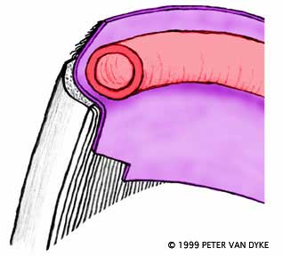 The coronary corium is the tissue that produces the horn that makes up the hoof wall. Its blood is supplied through the coronary plexus above it. The coronary plexus is drawn in this cross section as a "tube" for illustrative purposes. The real plexus is more like a sponge than a tube. The coronary corium is the tissue that produces the horn that makes up the hoof wall. Its blood is supplied through the coronary plexus above it. The coronary plexus is drawn in this cross section as a "tube" for illustrative purposes. The real plexus is more like a sponge than a tube.
move your mouse over the illustration
The dorsal aspect of the tube is compressed or "pinched" by the tissues descending into it. The amount of blood flow processed through the coronary plexus to the coronary corium in the pinched area will be less than in areas where it is not pinched. When blood flow is normal along the length of the coronary plexus the coronary corium will produce horn at an equal rate along its length. When blood flow is inhibited along the dorsal region of the coronary plexus the coronary corium will produce horn at a slower rate in the dorsal region.
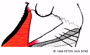 The "dish" is the product of unequal growth rates along the length of the coronary corium. The dorsal region is growing horn slowly when that quarters and heels are growing at a faster rate. The heels outgrow the toe. The "bumps" at the toe and lines along the heel represent growth rings or "fever rings". You can see that the distance between the "rings" increases as they approach the heel. The "dish" is the product of unequal growth rates along the length of the coronary corium. The dorsal region is growing horn slowly when that quarters and heels are growing at a faster rate. The heels outgrow the toe. The "bumps" at the toe and lines along the heel represent growth rings or "fever rings". You can see that the distance between the "rings" increases as they approach the heel.
You can move your mouse over this illustration, but not much will happen!

© Copyright MMII Peter Van Dyke
|