
|
Radiograph Preparation
|
Radiographs are used to evaluate hoof anatomy and to track therapeutic progress (or lack of progress). Because radiographs do not image horn very well radiopaque markers are placed on the outside of the hoof to indicate those oocations on the radiograph.
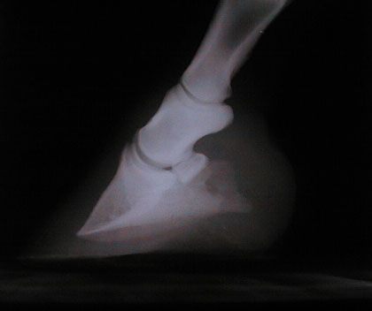
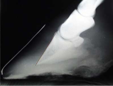
Move your mouse over the image. These two images were made within minutes of one another. No markers were used in the first image. The mouse-over image was made using a stage with a copper wire imbeded into its surface. A 60mm stainless steel rod was taped along the center of the toe with its upper end placed on the palpable skin/horn junction.
Founder Distance is the most useful measurement in a lateral view. Founder Distance (FD) is the difference in height between the skin/hair junction and the extensor process. This is a measurement not suseptable to changes made by trimming or otherwise altering the exterior of the hoof capsule.
We identify three locations on the radiograph; the skin/horn junction at the center of the toe (sagittally), the junction of the "live" frog and sole (not shown in images above), and a baseline (horizon) for the radiograph. Hair Line and Wall Profile The skin/horn junction is marked with a metallic rod taped to the center of the hoof wall. Palpation of the hair line will reveal the location of the skin/horn junction. Place the top of the rod on the skin/horn junction at the center of the hoof. Note how you locate this spot so that you can place the top of the rod at the same location in subsequent radiographs. The rod should be of known length to enable calibration of the radiograph for measurement purposes. We use 2.5mm stainless steel rods 60mm in length. Frog Locate the junction of the frog and sole at the apex of the frog and place a marker there. We use this point because it is an easily located landmark on the sole. If this point is tender the marker may be placed a known distance (10mm) behind this point. Note how marker placement was made so it can be repeated in future radiographs. This point may be transferred mediolaterally to the walls using a straight edge and notches made to where the straight edge crosses the walls. This allows us to identify the point on the sole again should the original marker be lost. Baseline To mark the horizontal baseline embed a 2mm copper or stainless wire along the top surface of the radiograph stage or block under the midpoint of where the hoof will rest on the stage. The radiograph machine must be placed such that the beam it generates will be both perpendicular and parallel to the hoof at the height of the hair line at the toe. The film focal distance and object film distance should be noted for future reference and for maintaining consistency between films. How the hoof is trimmed will affect measurements involving external surfaces. This should be kept in mind when trimming the feet prior to making radiographs. A methodological approach to preparation of hooves for laminitis/founder radiographic evaluation will produce higher quality and consistency. When possible trim the hoof for subsequent radiographs as it was trimmed for the original radiograph. Photographs mimicking the radiographs should also be made. A photograph taken with the same care from the same perspective as the radiograph machine should be taken immediately before or after the radiograph is made. This will record the radiograph photographically providing valuable information not often recorded.
Move your mouse over the image.

|
© Copyright MMII Peter Van Dyke