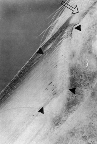
Normal Coronary Papillae |
|
On mouse over:
|
|
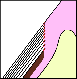
Photo courtesy of RA Eustace, p5 Explaining Laminitis and its Prevention by Robert A. Eustace MRCVS Available from: Life Data Labs http://www.lifedatalabs.com/ The main tissue types have been colored just to give a general idea of their locations. For a detailed discussion of hoof capsule histology visit Dr. Pollitt's web pages: http://www.uq.edu.au/~apcpolli (click through "proceed" and go to "publications/articles").
Note the parallelism between the hoof wall and P3, orientation of the coronary papillae and the density of the coronary papillae. There are two important watersheds associated with laminitis and founder. Without benefit of therapy laminitis will follow a course either toward recovery or toward founder, and founder will follow a course either toward chronic founder or toward death (see note on death). These two watersheds identify four pathways. Each path has differing therapeutic requirements. The purpose of therapy is to direct the course of the disease along a specific pathway. Generally speaking a therapeutic program designed to influence laminitis cases toward recovery and away from founder is not recommended as a therapeutic program capable of directing a founder cases toward chronic founder and away from death. When selecting a therapy it is essential that these watersheds and their resulting pathways are kept in mind and that selected therapies are capable of directing the disease to the desired conclusion. Understand the pathology and its potential outcomes. And understand therapeutic options and their capacities to favorably affect the pathology. |

© Copyright MMII Peter Van Dyke